List the three basic functions of the vertebrate kidney. Describe the adaptations in terrestrial animals to cope with water loss.
Functions of the vertebrate kidney
The three basic functions of the vertebrate kidney are (i) Filtration (ii) Reabsorption (iii) Secretion.
The adaptations in terrestrial animals to cope with water loss In the terrestrial environment, animal faces the problem of loss of body water due to the arid condition. They survive in arid conditions by some following adaptations:-
(i) Water movement through the integument: The integument of most terrestrial animal is relatively impermeable to water & very little water is lost through the skin. For example, insects lose very little moisture through the integument. It is due to the presence of waxy cuticle which is highly impermeable to water.
(ii) Water loss during air breathing: Water is lost through the respiratory surface. In the terrestrial vertebrates, the evaporative loss is reduced because the respiratory surface in them (the lungs) is internal to the body cavity. In a number of vertebrates, the respiratory loss of water is minimized through a mechanism known as a temporal counter current system. And the major route of water loss in terrestrial insects is through tracheal system.
(iii) Water loss during excretion: In terrestrial animals, body water is also lost during excretion of nitrogenous wastes. In terrestrial vertebrates, the kidney is the chief organ of osmoregulation & excretion.
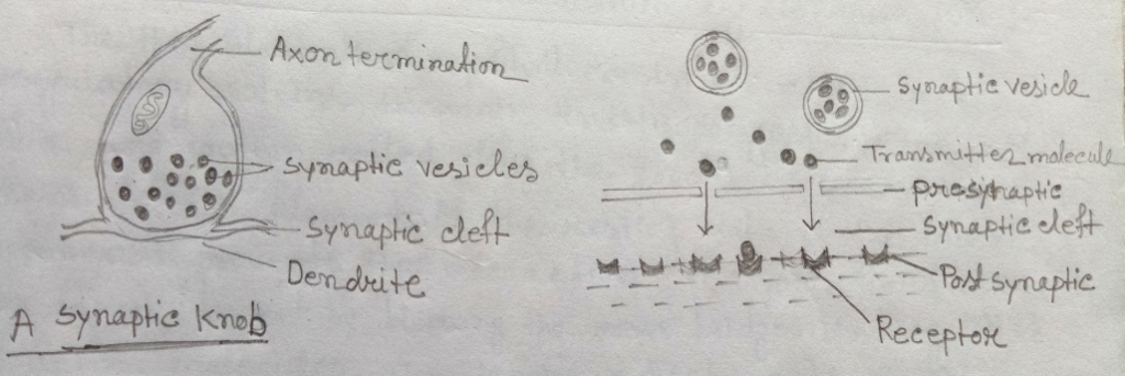
b) Explain synaptic transmission at the neuromuscular junction with the help of a suitable diagram.

Fig: Synaptic transmission
Synaptic transmission of information is mostly through chemical synapses & in some instances through electrical synapses. Chemical synapses involve neurotransmitter substances that are produced in presynaptic terminals & released in the narrow cleft that separates the presynaptic cell from postsynaptic cell. Receptors in the postsynaptic membrane pick up these transmitter molecules. These receptors are linked to chemically gated ion channels & change in membrane permeability generates a postsynaptic potential which may be inhibitory or excitatory. In excitatory postsynaptic potential, the action of the transmitter molecules is to shift the membrane potential beyond the threshold for initiation of the action potential in the axon hillock. Inhibitory postsynaptic potential changes in conduction so as to counter act depolarization of the membrane.
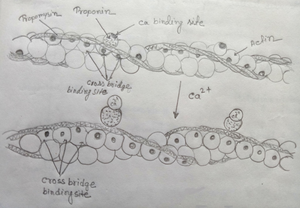
c) Discuss how calcium ions act as physiological regulators of muscle contraction.
The thin filaments of myofibril consist of action & regulatory proteins, troponin & tropomyosin. Tropomyosin is a long protein coiled along the groove between the two chains of the actin filament. At the resting state of the muscle, tropomyosin prevents the interaction of myosin head with the actin filament by blocking the cross bridges binding sites of actin molecules. The tropin which acts as a controlling protein has an affinity for calcium ion. It has a binding site for Ca^2+. When a muscle a stimulated, the calcium ion concentration within the muscle fiber rises abruptly; the calcium ions bind to troponin & bring out conformational changes in both troponin & tropomyosin molecules. This effect induces the movement of tropomyosin to uncover the binding sites & allows cross bridges of the myosin to bind to the actin filament. Thus, calcium ion acts as the physiological regulator of muscle contraction.

Fig: Control of contraction by calcium ion