a) What does an oxygen dissociation curve show? Draw & compare the oxygen dissociation curves of hemoglobin & myoglobin.
The relationship of oxygen carrying capacity to surrounding oxygen concentration can be shown graphically by oxygen dissociation curves.
Comparison between the oxygen dissociation curves of hemoglobin & myoglobin

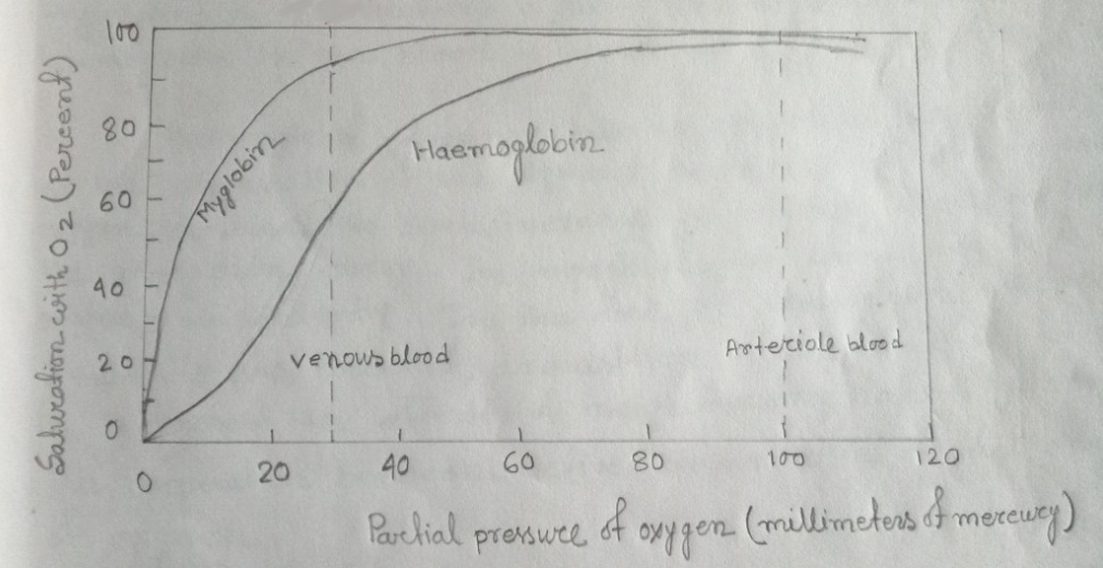
Fig: A comparison of the dissociation curves for hemoglobin & for myoglobin
The oxygen dissociation curve of hemoglobin is S-shaped or a sigmoid curve. This curve shows how hemoglobin's binding capacity depends on the partial pressure of oxygen & how hemoglobin acts as a carrier of oxygen. Also, the curve shows that changes in partial pressure (Po2) values from arterial to venous blood result in 97-75 = 22% unloading when resting. During exercise, this unloading is increased to 39%.
And the oxygen dissociation curve of myoglobin is rectangular. Myoglobin acts as a middleman in the transfer of oxygen from the blood to mitochondria within muscle cells. These curves indicate that the compound remains oxygenated until quite low levels of Po2 in the surrounding fluids is reached. According to the curves, at 20 mm Hg, the myoglobin in the muscles is above 80% saturated but the hemoglobin in the blood is about 30%.
Hemoglobin does not take up oxygen at low Po2 but as the oxygenation of the pigment occurs its affinity for more oxygen increases. No such increase in affinity is evident from the myoglobin curve. In myoglobin, the haem reacts to oxygen independently. In the case of hemoglobin where 4 subunits are present, acquisition of one molecule of O2 increases the affinity of neighboring haems for oxygen. This cooperation between active sites modifies the binding of oxygen.
b) Why do animals exhibit differences between their essential food requirements? Where does the absorption of carbohydrates, lipids & amino acids take place in the vertebrate body? Describe the method of glucose absorption
Animals exhibit differences between their essential food requirements because the ability to synthesize is genetic & species specific. Besides this, the feeding mechanism is also different. For instance, animals can synthesize vitamin C but human being can't synthesize vitamin C. Hence, the human being must take vitamin C to enhance immunity.
The absorption of carbohydrates, lipids & amino acids takes place in the small intestine.
The method of glucose absorption

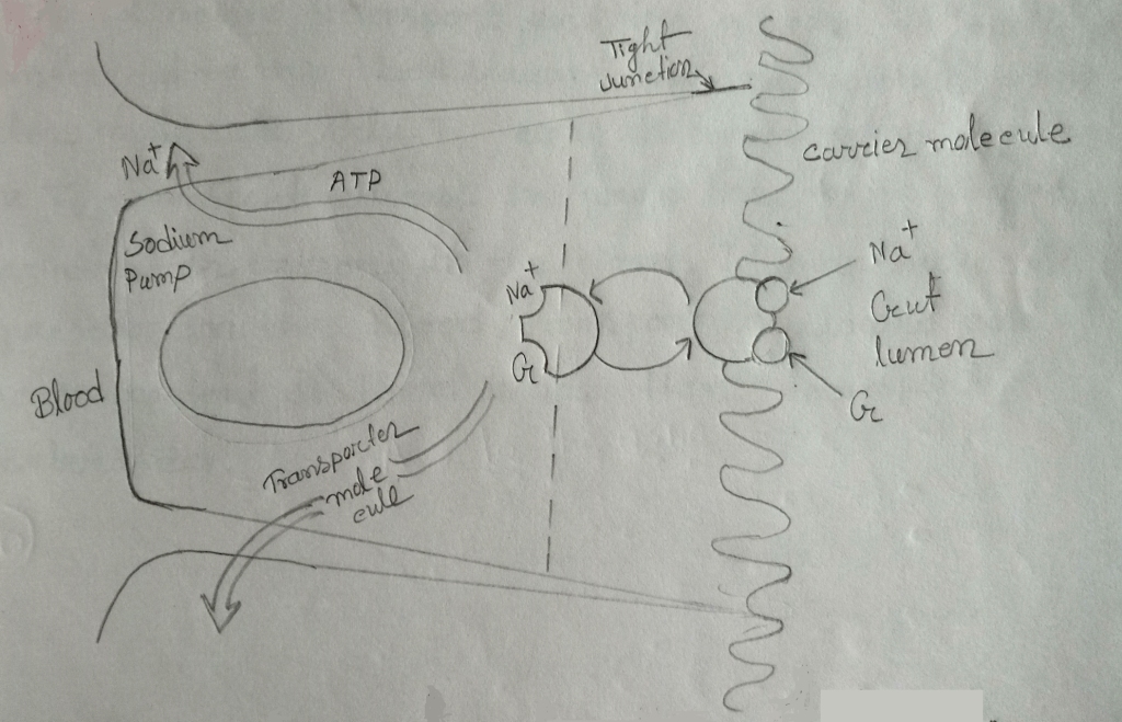
Fig: Mechanism for absorption of glucose
Sugars like glucose are absorbed through special transporter molecules in the membrane of the cell & depend on the Na^+ gradient between the lumen of the gut & cytoplasm of the epithelial cells. The molecule involved in absorption of glucose is known as cotransporter because it couples the transport of a glucose molecule with that of a sodium ion along its gradient. The cotransporter enables cells lining the lumen of the intestine to absorb even quite small traces of glucose from food even though the epithelial cells may already have high concentrations of glucose inside them.
Once inside the cell, the sodium ion is pumped out by ATP energized active transport & the glucose molecule is transferred to the blood stream through another transporter molecule, Glu T2, along with its concentration gradient. Glu T2 transport glucose in proportion to the sugar concentration present in the blood. If more glucose is present in the blood, transport is slowed & if glucose content of blood is low then transport is accelerated.